jueves, 27 de octubre de 2016

musculo Coracobrachialis

MUSCLE coracobrachialis
SHORT DESCRIPTION
  • Longus and flattened.
  • In half of their journey he is I is crossed by the musculocutaneous nerve.
  • Musculo also called perforated Casserius.
OCUPA
  • Located in the upper and medial part of the arm.
ORIGIN
  • medial side of the apex of the coracoid process (common tendon with the short head of the biceps).
INSERTION
  • anterior surface of the humerus superiorly to the middle and above the medial edge of the bone.
INNERVATION: Nerve Musculocutaneous.

FUNCTION:  previous Displaces and medially arm.
MUSCLE coracobrachialis

Shoulder muscles

SHOULDER MUSCLES
The shoulder muscles are divided into four groups:

PREVIOUS MUSCLE GROUP: They are arranged in two planes.

Deep
  1. Pectoral muscle Mayor
MUSCLE MEDIAL GROUP: C onstituido only by:
  1. Musculo Serrato Previous
BACK MUSCLE GROUP: comprising the muscles of the posterior wall of the axilla are in immediate relation to the scapula.
SIDE MUSCLE GROUP:  C onstituido only by:

Pectoral muscle Mayor

MAJOR MUSCLE CHEST
SHORT DESCRIPTION
  • Dorsi, flattened and triangular.
  • Thin medially and laterally thick.
  • It is separated from the deltoid muscle, forming the groove deltopectoral.
OCUPA
  • Located on the front and upper chest.
  • In front of the pectoralis minor.
  • It extends from the chest to the humerus.
ORIGIN: Portion:
  • Clavicular: 2/3 medial aspect of the anterior border of the clavicle. (Descending fibers)
  • Sternal: corresponding half of the sternum, 5-6 first costal cartilages. 4 and 5 are most important.
  • Abdominal: Front of the sheath of the rectus abdominis.
INSERTION
intertubercular anterior lip of the groove, using 2 fascicles:
  • Previous: Continuation of clavicular and sternal top of the fibers.
  • Rear: Formed by the other parts of the muscle.
These two issues are joined by means of a fascicle.

INNERVATION :  Nerves of the pectoralis major and minor, Brachial Plexus.

FUNCTION
  • Adductor and medial rotator arm.
  • fixed in the humeral point, raises the thorax. (Raising the whole body).

Minor Pectoral Muscle

Pectoralis MINOR
SHORT DESCRIPTION
  • lean, flat and triangular muscle.
  • It is covered by the pectoralis major muscle.
OCUPA
  • Located below the subclavian muscle. Forming a triangular space with this internal database called clavipectoral space.
ORIGIN
  • top edge and side face of the third, fourth and fifth ribs near the costal cartilages.
INSERTION
  • anterior medial border of the coracoid process, or the upper face of said process.
  • By a flattened tendon.
INNERVATION :  Nerve pectoralis minor, Brachial Plexus

FUNCTION
  • fixed point in the ribs, shoulder down the stump.
  • fixed point on the scapula, elevates the ribs (acting as inspiring muscle)


Subclavian muscle

MUSCLES SUBCLAVIAN
SHORT DESCRIPTION
  • Musculo elongated and fusiform.
OCUPA
  • Located below the collarbone.
  • It extends obliquely between the first rib and clavicle.
ORIGIN
  • upper face of the first costal cartilage and nearest part of the top face of the first rib.
  • By means of a cylindrical tendon.
INSERTION
  • underside of the clavicle, in an excavation groove called subclavian muscle.
INNERVATION :  Brachial Plexus

FUNCTION
  • fixed point on the first rib, clavicle lowered and therefore the shoulder.
  • collarbone fixed point, Eleva the first rib (acting as inspiring muscle).

Triangular muscle Sternum

MUSCLES TRIANGULAR breastbone
SHORT DESCRIPTION
  • Small muscle belly.
ORIGIN
  • In the xiphoid process and on the back side of the sternum.
INSERTION 
  • In the inner faces of the costal cartilages 2-6.
INNERVATION :  Intercostal nerves.

FUNCTION
  • They are differentiations of internal intercostal muscles.
  • They exist only in the intercostal spaces means (4, 5, 6, 7).
  • Saltan 2 or 3 ribs.
Triangular sternal muscles

Supracostales muscles

SUPRACOSTALES MUSCLES
SHORT DESCRIPTION
  • They are differentiations of the external intercostal muscles. They can be long or short.
ORIGIN
  • At the tip of the transverse process of a vertebra.
INSERTION
  • In the externosuperior infradyacente edge of the rib.
INNERVATION :  Intercostal nerves Correspondents.

FUNCTION
  • vertebral rotation: If the rib is set.
  • Lifting Rib: If the spine is fixed.
Supracostales muscles

Subcostal muscles

subcostal MUSCLES
SITUATION
  • Formed by the most posterior intercostal muscle fascicles intimate.
  • It extends from the inner side of a rib to the inner face of the underlying rib.
  • They exist only in the intercostal spaces means (4, 5, 6, 7). Saltan 2 or 3 ribs.
subcostal muscles

Ad

Minor serratus posterior inferior muscle

LOWER LOWER BACK MUSCLE SERRATO
SITUATION
  • It is located in the lower back.
  • It extends from the spine to the last 4 ribs.
ORIGIN
  • spinous process of the last two thoracic vertebrae and the first 3 lumbar vertebrae. As in the interspinous ligament.
  • Or in the spinous processes of T11-L2.
INSERTION
  • bottom edge and the side face of the 3 or 4 last ribs.
  • Or in the posterolateral aspect of the last 4 ribs.
INNERVATION : For the 9, 10 and 11 Intercostal Nerves.

FUNCTION
  • Lowering the ribs.
Serratus posterior inferior muscle
Ad

Minor serratus posterior superior muscle

UPPER LOWER BACK MUSCLE SERRATO
SITUATION
  • It is situated on the upper back.
  • It extends from the spine to the first ribs.
ORIGIN
  • Bottom of the nuchal ligament.
  • spinous processes of the seventh cervical vertebra and the first 3 thoracic vertebrae.
  • corresponding interspinous ligament.
INSERTION 
  • Lateral edge of the first rib.
  • side face and upper edge of the second, third and fourth rib.
  • Sometimes he reaches the fifth rib.
INNERVATION :  First Intercostals.

FUNCTION
  • Lift the ribs inspiring.
Serratus posterior superior muscle
Ad

Intimate intercostal muscle

Innermost intercostal muscle
SHORT DESCRIPTION
  • Medial internal intercostal muscle.
  • Its fibers descend obliquely lower and posteriorly.
ORIGIN
  • inner lip of the costal groove.
INSERTION
  • top edge of the underlying rib.
OCUPA
  • The intercostal region extending from the angle of the rib to 5 to 6 cm from the lateral edge of the sternum.
INNERVATION :  Intercostal nerves Correspondents.

FUNCTION
  • Rib tend to rise, pulling the upper raise the bottom. This occurs whenever the 1st or 2nd rib are fixed by the scalene.
OBSERVATION

  • Among the intimate intercostal muscle and internal intercostal muscle, you are the intercostal neurovascular bundle.

intimate intercostal muscle

Ad

Internal intercostal muscles

Intercostal muscles INMATES
SHORT DESCRIPTION
  • They observed by removing external intercostals.
  • Decienden fibers obliquely lower and posteriorly.
ORIGIN
  • Medially to the external intercostal M., along the outer apex of costal groove.
INSERTION
  • M. external intercostal medially along the top edge of the underlying rib.
OCUPA
  • The intercostal part between the mid-axillary line and the lateral edge of the sternum.
INNERVATION :  Intercostal nerves Correspondents.

FUNCTION
  • Expiration.
FACT 
  • Among the intimate intercostal muscle and internal intercostal muscle, you are the intercostal neurovascular bundle.
Internal intercostal muscles
Ad

External intercostal muscles

EXTERNAL intercostal muscles
SHORT DESCRIPTION
  • They are occupying the intercostal spaces and are facing downward and backward, leaving the latter part of the intercostal space.
  • They are 11 and there in the front.
  • The back specializes in a muscle called supracostal, which is fixed to the transverse processes of the thoracic vertebrae.
  • Their insertions occur through muscle and tendon fibers.
ORIGIN
  • outer lip of the costal groove of the upper rib intercostal space.
INSERTION
  • outer lip of the upper edge of the underlying rib.
OCUPA
  • then begins to costotransverse joint.
  • It was previously stopped in the vicinity of the costocondral joint, and continue with the external intercostal membrane.
INNERVATION :  Intercostal nerves Correspondents.

FUNCTION
  • Rib tend to rise, pulling the upper raise the bottom. This occurs whenever the 1st or 2nd rib are fixed by the scalene.
                                   EXTERNAL intercostal muscles
Ad

Serratus anterior muscle or

PREVIOUS OR MORE MUSCLE SERRATO
SHORT DESCRIPTION
  • Dorsi, flattened and quadrilateral.
OCUPA
  • It is applied to the lateral chest wall. It goes from the top 10 ribs to the medial border of the scapula.
ORIGIN: 3 servings:

First portion or above:
  • triangular face, which occupies the superomedial angle of the anterior surface of the scapula.
Second Portion Portion or Media:
  • anterior lip of the medial border of the scapula.
Third portion or lower portion:
  • triangular facet, which occupies the inferomedially part of the anterior surface of the scapula.
INSET: 3 servings:

First portion or above:
  • Earlier in the lateral edge of the first rib (inconstant).
  • external face of the second rib.
Second Portion Portion or Media:
  • outside of the 2nd, 3rd and 4th rib.
Third portion or lower portion:
  • outside of the 5th to the tenth rib. (Through six distinct fingerings).
INNERVATION :  Nerve Serrato Mayor (Bell Vein Ch.), Brachial Plexus.

FUNCTION
  • fixed point on the chest wall anteriorly and laterally displaces (rotational movement) scapula.
  • fixed point on the scapula, raises the rib (inspiring becomes).
Ad


Subscapularis muscle

MUSCLE subscapular
SHORT DESCRIPTION

Musculo thick, triangular and wide.

2 bursae are attached to this muscle:
  • subcoracoid bag.
  • subtendinous bag subscapularis muscle.
These two bags can communicate with each other and with the joint cavity.
There may be a third bag but is inconstant.

OCUPA
  • Located above the scapula and shoulder joint.
  • It extends from the subscapularis fossa to the upper end of the humerus.
ORIGIN
  • Entire length of the front face of the scapula, except near the shoulder joint area.
INSERTION
  • superomedial of the lesser tubercle and top of the ridge formed by the tuber.
INNERVATION :  Two or three born nerves of the brachial plexus

FUNCTION
  • medial rotation of the arm.
Ad

Supraspinatus

supraspinatus
SHORT DESCRIPTION
  • Musculo thick and has the shape of a triangular pyramid.
OCUPA
It extends from the supraspinatus fossa to the upper end of the humerus.
Goes below the:
  • acromioclavicular joint
  • And coracoacromial ligament
ORIGIN
  • ¾ of the supraspinatus medial fossa and the deep surface of the fascia that covers it.
INSERTION
  • superior facet of the greater tubercle.
INNERVATION : Nerve Suprascapular branch of the brachial plexus

FUNCTION
  • Raises the arm sliding laterally, it's abductor.
  • It acts as an active ligament of the shoulder joint.
Ad

Infraspinatus muscle

infraspinatus muscle
SHORT DESCRIPTION
  • flat and triangular muscle
OCUPA
  • Located after the infraspinatus fossa and shoulder joint.
ORIGIN
  • Infraspinous fossa medially to the ridge bordering surfaces insertion of round muscles.
  • medial third of the infraspinatus fascia.
  • fibrous septum that separates it from major and minor round.
INSERTION
  • half a page of the greater tubercle.
INNERVATION : Nerve Suprascapular branch of the brachial plexus.

FUNCTION
  • abductor and lateral rotator arm.
  • Contact helps keep the shoulder joint surface. 
Ad

Minor teres

Teres minor muscle
SHORT DESCRIPTION
  • It is a band-like muscle, elongated scalloping (va making an "S"). It gets its name because the visible part gives a rounded appearance, the rest is covered by the deltoid.
OCUPA
  • Located immediately below the infraspinatus and post M. shoulder joint.
ORIGIN
  • upper axillary border of the scapula (infraspinatous fossa).
INSERTION
  • lower facet of the greater tubercle.
INNERVATION : Nerve Circumflex

FUNCTION
  • abductor and lateral rotator arm.
Ad

Mayor teres

Teres MAYOR
SHORT DESCRIPTION
  • His name is given because of the rounded appearance of the visible portion presents is covered by other muscle mass as the latissimus dorsi and triceps.
OCUPA
  • Located below the M. teres minor.
ORIGIN
  • inferiorolateral part of the infraspinatus fossa. Laterally inserting the infraspinatus muscle and lower the round M. greater.
INSERTION
  • intertubercular medial lip of the groove.
INNERVATION : Nerve Brachial Plexus Mayor Redondo

FUNCTION
  • adductor and medial rotator arm.
  • fixed in the humeral point, raises the inferior angle of the scapula and shoulder stump.

latissimus dorsi

Latissimus dorsi
SHORT DESCRIPTION
  • Dorsi, flat and thin.
  • Covers the back and bottom of the trunk.
  • It helps to form the back wall of the axillary region.
OCUPA
  • It extends from the thoracic, lumbar and sacral regions of the spine, to the intertubercular groove of the humerus.
ORIGIN
  • Prickly and the supraspinal ligaments in the last 6 thoracic vertebrae and five lumbar vertebrae process.
  • median sacral crest.
  • posterior third of the iliac crest.
  • external face of the last 4 ribs.
  • On its way it can give an accessory fasciculus, which goes to the inferior angle of the scapula.
INSERTION
  • Intertubercular bottom groove.
INNERVATION :  For the Fifth Cervical.

FUNCTION
  • Then pulls the arm, and gives a medial rotation movement.
  • Abduction.
  • Extension.
  • fixed in the humeral point, raises the trunk.
  • If you have 2 fixed ends: position contributes to the firm.
Ad

Deltoid muscle

MUSCLE ANATOMY DELTOIDES
SHORT DESCRIPTION
  • Shaped hollow half-cone with the base situated superiorly and vertex at the bottom.
  • It is the muscle that forms the shoulder stump.
OCUPA
  • side of the shoulder.
ORIGIN
  • 1/3 side of the front edge of the clavicle.
  • Vertex and lateral edge of the acromion.
  • lower side of the rear edge of the spine of the scapula.
INSERTION
  • lateral aspect of the middle third of the humerus, the deltoid tuberosity in or V deltoid.
INNERVATION: Nerve Circumflex

FUNCTION
  • Flexion - extension.
  • Abduction - adduction.
  • external - internal rotation.
Ad

bone hydrogen peroxide bleaching (osteotecnia)

UNIVERSITY ANDINA CUSCO
FACULTY OF HEALTH SCIENCES
CAREER OF MEDICINE HUMAN
Bleached bones Osteotecnia
BLEACHING BONE IN PEROXIDE  HYDROGEN  (OSTEOTECNIA)

INTRODUCTION
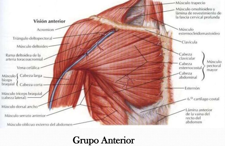
The teaching of human medicine is faced different Institutions With anatomical pieces Obtaining biological as teaching material. For This reason, it is of utmost Importance, knowledge of the preservation and restoration of materials present in laboratories, using accessible techniques and low cost.
The Osteotecnia, Refers to the various techniques for the preparation and preservation of bone pieces, Either for use in educational anatomy labs or for demonstration purposes, as it is in museums. It is based on the collection Primarily, cleaning and bleaching of bone material.

FRAMEWORK THEORETICAL
Composition of bone
Our bones are made of a materials called bone tissue. This fabric has the distinction of being very hard That Comprise Because the cells are surrounded by it hard minerals like calcium and phosphorus. Also it found in tissue esta resistant protein called collagen.
  •  40% - organic matrix. Mainly collagen type I; It contains 20% water
  •  60% -. Inorganic compounds different types of calcium salts as the body decomposes, let's go find us With less organic matter, Until It Disappears completely.
Other Things That can be found:
  •      Remains of tendons, cartilage, etc.
  •      Earth.
THEREFORE we will meet several "types" of bones, And This will depend on the depth of cleaning required:

  1. Bones With abundant organic matter:  Generally dry, it is Recognized by ITS darker colored, with respect to the bone; May Represent a tendon, cartilage, periosteum a thin film of brown Easily it follows bone, etc.
  2. With Bones Total absence of organic matter:  They Have a whitish in color; In These elapsed time required for complete decomposition of the organic remains, Therefore Comprised They are purely by inorganic materials (calcium salts and other minerals).
Obviously, Between 1 and 2 are all imaginable shades.

How does bleaching bones?
In cleaning, Most Often With traces of bones soft tissues, Which are removed with a knife Carefully, not to cause scratches or scrapes on the surface are acquired. Another option would expect the putrefaction process destroys the tissue, however much time is longer.
You May still adhere muscle, cartilage, earth elements, Therefore, a technique called maceration, Which is used to remove waste and processed to give a white appearance is applied.
The bone pieces can be boiled in water with soap, rinsed with water and then a drying allowed outdoors, under the sun or Either in shade.
With more time available, parts in water, Wherein the soft materials will lose its consistency detaching and Immersed.
In the end it is Easily removed using brushes and hot water.
It is Advisable to choose fresh corpses, Those Who Have Given That much time to be impregnated With formalin, brittle and likely to break When handled in cleaning.
After cleaning, some bones left residual With dark colored spots or a THROUGHOUT the piece.
This should not be a problem since it is possible to get a bone whitening Desired.

OBJECTIVES

GENERAL OBJECTIVE:
·          Conservation of bone material.
SPECIFIC OBJECTIVES:
· To          study and Implement the Osteotecnia in bone pieces.
·          Using chemical methods for whitening bone pieces .

MATERIALS
  • 2 tubs, you can enter in Which parts Hosea. It is advised That are longer than deep.
  • Detergent.
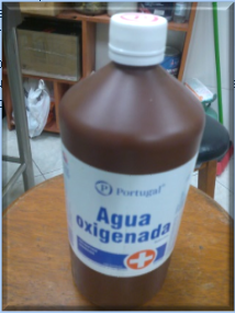
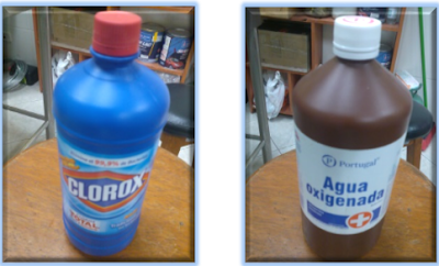
  • Hydrogen Peroxide 3% (peroxide).
  • 3% chlorine.
  • Whisk broom.
  • Water paper scissors (optional)
SCHEDULE
ACTIVITIES
CALENDAR
Obtaining bone pieces
9 - March 11, 2015
Obtaining materials
March 12, 2015
performance
March 14, 2015
ends
When bleaching is Achieved Desired
DESAROOLLO OF BLEACHING BONE IN HYDROGEN PEROXIDE
The bone pieces That Might get are:
PART BONE
STATE
humerus
With very little organic matter.
Radio
Varnished.
ulna
Painted silver.
Being in different states bone pieces will proceed to apply the technique to 3 and see the results.

First step, March 14, 2015
Materials:
  • bone pieces
  • 1 tub
  • Detergent
The method was applied to degrease the samples.
  • Prepare the detergent in the tub with water, making good foaming.
  • Immerse the bone pieces are Until They covered.                
       
This should be soaked for 1 week, to Achieve good degreasing.

Step Friday March 21, 2015

Materials:
  • Whisk broom
  • Scissors
  • bone pieces
  • 1 tub
  • (peroxide  hydrogen )
    After leaving to soak for a week we proceed to :
  • Remove fat and small traces of organic kill the humerus.
  • In the case of Radio it is varnished, and will be at the point of being reliable to make the varnish.
  • In the case of the ulna, the painting can get Already with the help of scissors
    Bleached bones OsteotecniaBleached bones Osteotecnia             Bleached bones OsteotecniaBleached bones Osteotecnia
For you can use the esta clothes brush, scissors or water sandpaper, Very Carefully not to damage the anatomical accidents. To Achieve a good cleaning.
Prepare the H 2 O 2

The preparation of peroxide  hydrogen  to water ratio in the range of 1 to 2 then:

If I put 1 ml of H 2 O 2 I will 2ml of H 2 O.

Using relationship for the preparation esta.
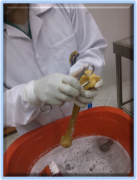
  • For bleaching in my case using 800 ml of H and 1600 ml of H O, This amount was to Achieve immersing the bone pieces.
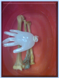
  • Let soak for 24 hrs.
Bleached bones Osteotecnia
Peroxide bleached bones to penetrate it, and molecules react With That cause spots or discoloration. Generally, the higher the amount of peroxide, the greater the whitening power.
Bleached bones OsteotecniaBleached bones Osteotecnia

The humeral head can reach your weight float esta That presents, so it can be used to Achieve a weight dipping as in the image.

Step Monday March 23, 2015

Materials:

  • bony parts
  • 2 tubs
  • 2
  • Bleach

Bleached bones Osteotecnia
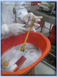
The preparation of the bleach with water will be in the ratio of 1 ml of water 5 ml bleach.

To Achieve my case and submerging the bone pieces to Prepare:
  • 500 ml  of liquor in 2500 ml of H O
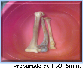
After leaving for 24Hrs peroxide  hydrogen  With The bones will proceed to change container every 5 minutes.

That Means that:
  • 5 min.  In the preparation of bleach
  • 5 min.  In the preparation of H 2
Bleached bones Osteotecnia       Bleached bones Osteotecnia
This process must be Repeated Several times Desired Until the target.
Be very careful in the days When the bones are left in the bleach as decalcified These parts and can crack or break reach in the worst case.
If it was not possible on the day Indicated the bone whitening , you can leave the bones in the H 2 O 2 , but not in the bleach from the above explanation.

WORK COMPLETED
ad
TO THE PRESENTATION:
Materials:
  • Wood
  • Suspenders


   Presentation:

Bibliography:

Dr. Concha I. Techniques Anatomic. [Internet]. 2nd ed. Anato.cl. 2007. [updated 15sep 2011; cited 14 March 2015]. Available at: http: // www.anato.cl/etecnica/clasesvideos/Sylltecanat06.pdf
MSc. Sanches G. Cañete M. Craft Assembling a skeleton mediantes osteotecnia variants. [Internet]. Volume 15 No. 09. REDVET. 2014. [Cited 14 March 2015]. Available in: http://www.veterinaria.org/revistas/redvet/n090914/091410.pdf