Mostrando entradas con la etiqueta Anatomy. Mostrar todas las entradas

jueves, 27 de octubre de 2016

musculo Coracobrachialis

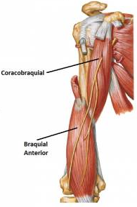
MUSCLE coracobrachialis
SHORT DESCRIPTION
  • Longus and flattened.
  • In half of their journey he is I is crossed by the musculocutaneous nerve.
  • Musculo also called perforated Casserius.
OCUPA
  • Located in the upper and medial part of the arm.
ORIGIN
  • medial side of the apex of the coracoid process (common tendon with the short head of the biceps).
INSERTION
  • anterior surface of the humerus superiorly to the middle and above the medial edge of the bone.
INNERVATION: Nerve Musculocutaneous.

FUNCTION:  previous Displaces and medially arm.
MUSCLE coracobrachialis

Shoulder muscles

SHOULDER MUSCLES
The shoulder muscles are divided into four groups:

PREVIOUS MUSCLE GROUP: They are arranged in two planes.

Deep
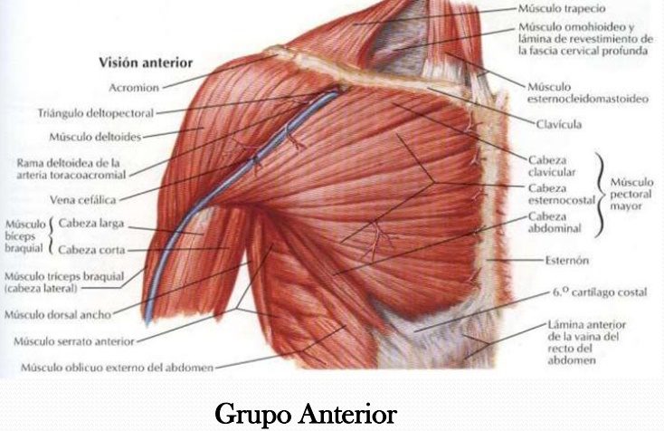
  1. Pectoral muscle Mayor
MUSCLE MEDIAL GROUP: C onstituido only by:
  1. Musculo Serrato Previous
BACK MUSCLE GROUP: comprising the muscles of the posterior wall of the axilla are in immediate relation to the scapula.
SIDE MUSCLE GROUP:  C onstituido only by:

Pectoral muscle Mayor

MAJOR MUSCLE CHEST
SHORT DESCRIPTION
  • Dorsi, flattened and triangular.
  • Thin medially and laterally thick.
  • It is separated from the deltoid muscle, forming the groove deltopectoral.
OCUPA
  • Located on the front and upper chest.
  • In front of the pectoralis minor.
  • It extends from the chest to the humerus.
ORIGIN: Portion:
  • Clavicular: 2/3 medial aspect of the anterior border of the clavicle. (Descending fibers)
  • Sternal: corresponding half of the sternum, 5-6 first costal cartilages. 4 and 5 are most important.
  • Abdominal: Front of the sheath of the rectus abdominis.
INSERTION
intertubercular anterior lip of the groove, using 2 fascicles:
  • Previous: Continuation of clavicular and sternal top of the fibers.
  • Rear: Formed by the other parts of the muscle.
These two issues are joined by means of a fascicle.

INNERVATION :  Nerves of the pectoralis major and minor, Brachial Plexus.

FUNCTION
  • Adductor and medial rotator arm.
  • fixed in the humeral point, raises the thorax. (Raising the whole body).

Minor Pectoral Muscle

Pectoralis MINOR
SHORT DESCRIPTION
  • lean, flat and triangular muscle.
  • It is covered by the pectoralis major muscle.
OCUPA
  • Located below the subclavian muscle. Forming a triangular space with this internal database called clavipectoral space.
ORIGIN
  • top edge and side face of the third, fourth and fifth ribs near the costal cartilages.
INSERTION
  • anterior medial border of the coracoid process, or the upper face of said process.
  • By a flattened tendon.
INNERVATION :  Nerve pectoralis minor, Brachial Plexus

FUNCTION
  • fixed point in the ribs, shoulder down the stump.
  • fixed point on the scapula, elevates the ribs (acting as inspiring muscle)


Subclavian muscle

MUSCLES SUBCLAVIAN
SHORT DESCRIPTION
  • Musculo elongated and fusiform.
OCUPA
  • Located below the collarbone.
  • It extends obliquely between the first rib and clavicle.
ORIGIN
  • upper face of the first costal cartilage and nearest part of the top face of the first rib.
  • By means of a cylindrical tendon.
INSERTION
  • underside of the clavicle, in an excavation groove called subclavian muscle.
INNERVATION :  Brachial Plexus

FUNCTION
  • fixed point on the first rib, clavicle lowered and therefore the shoulder.
  • collarbone fixed point, Eleva the first rib (acting as inspiring muscle).

Triangular muscle Sternum

MUSCLES TRIANGULAR breastbone
SHORT DESCRIPTION
  • Small muscle belly.
ORIGIN
  • In the xiphoid process and on the back side of the sternum.
INSERTION 
  • In the inner faces of the costal cartilages 2-6.
INNERVATION :  Intercostal nerves.

FUNCTION
  • They are differentiations of internal intercostal muscles.
  • They exist only in the intercostal spaces means (4, 5, 6, 7).
  • Saltan 2 or 3 ribs.
Triangular sternal muscles

Supracostales muscles

SUPRACOSTALES MUSCLES
SHORT DESCRIPTION
  • They are differentiations of the external intercostal muscles. They can be long or short.
ORIGIN
  • At the tip of the transverse process of a vertebra.
INSERTION
  • In the externosuperior infradyacente edge of the rib.
INNERVATION :  Intercostal nerves Correspondents.

FUNCTION
  • vertebral rotation: If the rib is set.
  • Lifting Rib: If the spine is fixed.
Supracostales muscles

Subcostal muscles

subcostal MUSCLES
SITUATION
  • Formed by the most posterior intercostal muscle fascicles intimate.
  • It extends from the inner side of a rib to the inner face of the underlying rib.
  • They exist only in the intercostal spaces means (4, 5, 6, 7). Saltan 2 or 3 ribs.
subcostal muscles

Ad

Minor serratus posterior inferior muscle

LOWER LOWER BACK MUSCLE SERRATO
SITUATION
  • It is located in the lower back.
  • It extends from the spine to the last 4 ribs.
ORIGIN
  • spinous process of the last two thoracic vertebrae and the first 3 lumbar vertebrae. As in the interspinous ligament.
  • Or in the spinous processes of T11-L2.
INSERTION
  • bottom edge and the side face of the 3 or 4 last ribs.
  • Or in the posterolateral aspect of the last 4 ribs.
INNERVATION : For the 9, 10 and 11 Intercostal Nerves.

FUNCTION
  • Lowering the ribs.
Serratus posterior inferior muscle
Ad

Minor serratus posterior superior muscle

UPPER LOWER BACK MUSCLE SERRATO
SITUATION
  • It is situated on the upper back.
  • It extends from the spine to the first ribs.
ORIGIN
  • Bottom of the nuchal ligament.
  • spinous processes of the seventh cervical vertebra and the first 3 thoracic vertebrae.
  • corresponding interspinous ligament.
INSERTION 
  • Lateral edge of the first rib.
  • side face and upper edge of the second, third and fourth rib.
  • Sometimes he reaches the fifth rib.
INNERVATION :  First Intercostals.

FUNCTION
  • Lift the ribs inspiring.
Serratus posterior superior muscle
Ad

Intimate intercostal muscle

Innermost intercostal muscle
SHORT DESCRIPTION
  • Medial internal intercostal muscle.
  • Its fibers descend obliquely lower and posteriorly.
ORIGIN
  • inner lip of the costal groove.
INSERTION
  • top edge of the underlying rib.
OCUPA
  • The intercostal region extending from the angle of the rib to 5 to 6 cm from the lateral edge of the sternum.
INNERVATION :  Intercostal nerves Correspondents.

FUNCTION
  • Rib tend to rise, pulling the upper raise the bottom. This occurs whenever the 1st or 2nd rib are fixed by the scalene.
OBSERVATION

  • Among the intimate intercostal muscle and internal intercostal muscle, you are the intercostal neurovascular bundle.

intimate intercostal muscle

Ad

Internal intercostal muscles

Intercostal muscles INMATES
SHORT DESCRIPTION
  • They observed by removing external intercostals.
  • Decienden fibers obliquely lower and posteriorly.
ORIGIN
  • Medially to the external intercostal M., along the outer apex of costal groove.
INSERTION
  • M. external intercostal medially along the top edge of the underlying rib.
OCUPA
  • The intercostal part between the mid-axillary line and the lateral edge of the sternum.
INNERVATION :  Intercostal nerves Correspondents.

FUNCTION
  • Expiration.
FACT 
  • Among the intimate intercostal muscle and internal intercostal muscle, you are the intercostal neurovascular bundle.
Internal intercostal muscles
Ad

External intercostal muscles

EXTERNAL intercostal muscles
SHORT DESCRIPTION
  • They are occupying the intercostal spaces and are facing downward and backward, leaving the latter part of the intercostal space.
  • They are 11 and there in the front.
  • The back specializes in a muscle called supracostal, which is fixed to the transverse processes of the thoracic vertebrae.
  • Their insertions occur through muscle and tendon fibers.
ORIGIN
  • outer lip of the costal groove of the upper rib intercostal space.
INSERTION
  • outer lip of the upper edge of the underlying rib.
OCUPA
  • then begins to costotransverse joint.
  • It was previously stopped in the vicinity of the costocondral joint, and continue with the external intercostal membrane.
INNERVATION :  Intercostal nerves Correspondents.

FUNCTION
  • Rib tend to rise, pulling the upper raise the bottom. This occurs whenever the 1st or 2nd rib are fixed by the scalene.
                                   EXTERNAL intercostal muscles
Ad

Serratus anterior muscle or

PREVIOUS OR MORE MUSCLE SERRATO
SHORT DESCRIPTION
  • Dorsi, flattened and quadrilateral.
OCUPA
  • It is applied to the lateral chest wall. It goes from the top 10 ribs to the medial border of the scapula.
ORIGIN: 3 servings:

First portion or above:
  • triangular face, which occupies the superomedial angle of the anterior surface of the scapula.
Second Portion Portion or Media:
  • anterior lip of the medial border of the scapula.
Third portion or lower portion:
  • triangular facet, which occupies the inferomedially part of the anterior surface of the scapula.
INSET: 3 servings:

First portion or above:
  • Earlier in the lateral edge of the first rib (inconstant).
  • external face of the second rib.
Second Portion Portion or Media:
  • outside of the 2nd, 3rd and 4th rib.
Third portion or lower portion:
  • outside of the 5th to the tenth rib. (Through six distinct fingerings).
INNERVATION :  Nerve Serrato Mayor (Bell Vein Ch.), Brachial Plexus.

FUNCTION
  • fixed point on the chest wall anteriorly and laterally displaces (rotational movement) scapula.
  • fixed point on the scapula, raises the rib (inspiring becomes).
Ad


Subscapularis muscle

MUSCLE subscapular
SHORT DESCRIPTION

Musculo thick, triangular and wide.

2 bursae are attached to this muscle:
  • subcoracoid bag.
  • subtendinous bag subscapularis muscle.
These two bags can communicate with each other and with the joint cavity.
There may be a third bag but is inconstant.

OCUPA
  • Located above the scapula and shoulder joint.
  • It extends from the subscapularis fossa to the upper end of the humerus.
ORIGIN
  • Entire length of the front face of the scapula, except near the shoulder joint area.
INSERTION
  • superomedial of the lesser tubercle and top of the ridge formed by the tuber.
INNERVATION :  Two or three born nerves of the brachial plexus

FUNCTION
  • medial rotation of the arm.
Ad

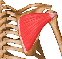
Supraspinatus

supraspinatus
SHORT DESCRIPTION
  • Musculo thick and has the shape of a triangular pyramid.
OCUPA
It extends from the supraspinatus fossa to the upper end of the humerus.
Goes below the:
  • acromioclavicular joint
  • And coracoacromial ligament
ORIGIN
  • ¾ of the supraspinatus medial fossa and the deep surface of the fascia that covers it.
INSERTION
  • superior facet of the greater tubercle.
INNERVATION : Nerve Suprascapular branch of the brachial plexus

FUNCTION
  • Raises the arm sliding laterally, it's abductor.
  • It acts as an active ligament of the shoulder joint.
Ad

Infraspinatus muscle

infraspinatus muscle
SHORT DESCRIPTION
  • flat and triangular muscle
OCUPA
  • Located after the infraspinatus fossa and shoulder joint.
ORIGIN
  • Infraspinous fossa medially to the ridge bordering surfaces insertion of round muscles.
  • medial third of the infraspinatus fascia.
  • fibrous septum that separates it from major and minor round.
INSERTION
  • half a page of the greater tubercle.
INNERVATION : Nerve Suprascapular branch of the brachial plexus.

FUNCTION
  • abductor and lateral rotator arm.
  • Contact helps keep the shoulder joint surface. 
Ad

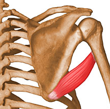
Minor teres

Teres minor muscle
SHORT DESCRIPTION
  • It is a band-like muscle, elongated scalloping (va making an "S"). It gets its name because the visible part gives a rounded appearance, the rest is covered by the deltoid.
OCUPA
  • Located immediately below the infraspinatus and post M. shoulder joint.
ORIGIN
  • upper axillary border of the scapula (infraspinatous fossa).
INSERTION
  • lower facet of the greater tubercle.
INNERVATION : Nerve Circumflex

FUNCTION
  • abductor and lateral rotator arm.
Ad

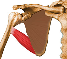
Mayor teres

Teres MAYOR
SHORT DESCRIPTION
  • His name is given because of the rounded appearance of the visible portion presents is covered by other muscle mass as the latissimus dorsi and triceps.
OCUPA
  • Located below the M. teres minor.
ORIGIN
  • inferiorolateral part of the infraspinatus fossa. Laterally inserting the infraspinatus muscle and lower the round M. greater.
INSERTION
  • intertubercular medial lip of the groove.
INNERVATION : Nerve Brachial Plexus Mayor Redondo

FUNCTION
  • adductor and medial rotator arm.
  • fixed in the humeral point, raises the inferior angle of the scapula and shoulder stump.

latissimus dorsi

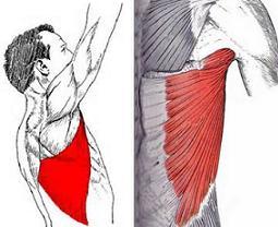
Latissimus dorsi
SHORT DESCRIPTION
  • Dorsi, flat and thin.
  • Covers the back and bottom of the trunk.
  • It helps to form the back wall of the axillary region.
OCUPA
  • It extends from the thoracic, lumbar and sacral regions of the spine, to the intertubercular groove of the humerus.
ORIGIN
  • Prickly and the supraspinal ligaments in the last 6 thoracic vertebrae and five lumbar vertebrae process.
  • median sacral crest.
  • posterior third of the iliac crest.
  • external face of the last 4 ribs.
  • On its way it can give an accessory fasciculus, which goes to the inferior angle of the scapula.
INSERTION
  • Intertubercular bottom groove.
INNERVATION :  For the Fifth Cervical.

FUNCTION
  • Then pulls the arm, and gives a medial rotation movement.
  • Abduction.
  • Extension.
  • fixed in the humeral point, raises the trunk.
  • If you have 2 fixed ends: position contributes to the firm.
Ad